
Instrument Snapshot
The Visualsonics Vevo F2 features an innovative signal pathway from transducer to screen that improves image clarity and frame rates.
In combination to its advanced technology for high-definition image processing, the Vevo F2 is ideal for cross-functional biological and physiological research in a wide range of animal models.
Vevo F2 offers flexibility to image at low frequency for penetration and ultra-high frequency for resolution using one platform (71–1 MHz).
Reach out to our core facility in Boston with any questions at cils-core@northeastern.edu.
Features
Linear probe: 57-25 MHz (UHF57x)
For high-resolution mouse cardiovascular, abdominal, reproductive; embryology; tumor imaging (< 14 mm diameter); small rat vascular
Linear probe: 29-15 MHz (UHF29x)
For rat cardiac & abdominal (<400 g); large tumor imaging (<23 mm diameter); all contrast applications
Phased probe: 8-4 MHz (p10xp)
For large animal cardiology & abdominal
Sample Images Acquired with Linear Probe
Application: Control Mouse – Short Axis Heart
Application: Control Mouse – Aortic Arch
Application: Control Mouse- Fully Elongated Heart (Annotated)
Rates
Internal NU
$60/h
External Academia
$110/h
Industry
$140/h
Visualsonics Vevo F2 Manual
VevoLab User Guide
FAQs
General Questions
What is Ultrasounds?
Ultrasounds (also called “sonography” or “ultrasonography”) is a non-invasive imaging technique that utilizes high-intensity sound waves to visualize soft tissue structures in real-time.
How does Ultrasounds work?
A transducer probe is placed on the area of interest with sufficient ultrasound gel to ensure proper transmission of the waves towards the tissue of interest. The ultrasounds probe converts electrical current into high-frequency sound waves, which bounce off different structures and are received back by the probe and converted into electrical signals again.
Imaging Questions
What is the resolution?
The image resolution will depend ultimately on the transducer probe utilized.
What do I need to be able to image an animal with Ultrasounds?
- Project approval by the director of the facility
- Safety training completed (BIOraft)
- Animal protocol approved (IACUC guidelines)
- MRI training by staff members of the facility successfully completed
- Reservation in FBS completed
How long is an Ultrasounds?
Ultrasounds image acquisitions are real-time, therefore images and videos can be acquired very fast. The major limiting factor in total study acquisition time is generally the preparation of the animal for imaging.
Animal-Related Questions
What animals or samples can I image with ultrasounds?
We currently have three transducer probes, that are specific to the animal size and organ of interest. Please contact us for more information.
Is there any lab space for animal preparation before or after the imaging study?
Yes, there is an adjacent room with available space for animal preparation as well as tissue collection procedures. Please discuss with the core director if you are planning on using these spaces for long periods of time (> 30 minutes) before or after the imaging experiments. These rooms MUST be included in the approved animal protocol before using them.
Is there a limit in sample/animal size? How big can my sample/animal be?
We currently have available a mouse platform, but normal sized rats can fit as well.
Does the animal need to be restricted during imaging?
Ideally, the animal should be as still as possible, to prevent any undesired artifacts in the image acquisition and facilitate positioning of the probe.
Do I need to bring my own anesthesia supply?
We offer inhaled isoflurane administered in a mixture of oxygen, in both the prep room and the imaging area.
Do I need to bring my own contrast agent for imaging?
Yes, users need to bring their own contrast agent, which MUST be included in the animal protocol.
Can I bring animals back to the animal housing facility?
Generally yes, but please discuss with DLAM before initiating your animal experiments for confirmation.
Do I need to follow IACUC guidelines?
Absolutely!! Users need to develop and amend their own IACUC protocol to include any imaging procedures in addition to room information. Descriptions of imaging methodologies and protocol templates are available upon request.
What should I add in the IACUC protocol?
For imaging studies without invasive procedures, we recommend adding a paragraph similar to the following example for mice in the Experimental Design section:
“The scope of this study will be limited to imaging of _____strain & animal____ without any invasive procedures. Ultrasounds is a non-invasive technique and generally regarded safe to animals. Imaging will be performed on the following parts of the animal: ______heart/brain/limbs/internal organs…____. No invasive procedure will be performed before or during the imaging
Mouse will be anesthetized with 1-3% isoflurane. Electrode gel will be applied to the EKG pads on the stage. Once anesthetized, the mouse will be transferred to the heated stage. Eye lubricant will be added to the eyes. Rectal probe will be lubricated with Aquagel lubricating gel before insertion. Heart rate, respiratory rate, and body temperature will be monitored. Nair will be applied with a cotton-tipped swab to remove hair from the head, chest, abdomen, and back. Nair will only be in contact with the animal for up to 1 minute to reduce skin irritation. Hair and nair will be removed with a wet sponge pad. Aquasonic clear ultrasound gel will be applied to the location of imaging (head, chest, abdomen, or back). Imaging will be performed with Visualsonics Vevo F2 Ultrasound for about 20 minutes. Time of preparation and imaging will take about 40 minutes for each mouse. After imaging is complete, ultrasound gel will be removed with wet sponge pads. Mouse will be monitored for recovery (ambulation, maintenance of sternal recumbency) from anesthesia before being returned to the animal facility. “
If surgery procedures or more advanced imaging protocols are needed, please contact us (g.rong@northeastern.edu) or DLAM (dlam@northeastern.edu)
Safety-Related Questions
Is Ultrasounds safe?
Ultrasounds is non-ionizing and under proper usage, it is safe/harmless for operators. Please discuss with the Core director if you have any safety concerns.
Will I be exposed to radiation or any side effects?
No- this instrument does not emit radiation and has no known side effects.
What can I (not) wear/have with me while at the Ultrasounds tfacility?
Lab coat, safety glasses, gloves (general PPE) is required.
Food and drinks are not permitted even in the control room.
Questions About Contrast Agents (CA)
What is an Ultrasounds contrast agent?
Ultrasounds contrast agents (UCA) are substances designed to facilitate the visualization of certain tissues of interest, commonly consisting of gas bubbles “microbubbles” encapsulated in different strategies for stabilization.
When is a contrast agent needed?
When alterations in the wave reflection pattern are needed to better identify or evaluate certain tissues or physiological mechanisms.
What are the risks of using contrast agents?
If the safety protocol and recommended guidelines are properly followed, there should be minimal risk to the operator associated with the use of Ultrasounds contrast agents.
When carrying animal studies consisting of contrast enhanced acquisitions, it is important to ensure that the dosage, administration route, and clearance considerations follow recommended guidelines and have received IACUC approval. Although minimal, there are some risks associated with improper injection of the contrast agent… If the operator is exposed to MRI contrast agents, please contact the CILS Core Director ASAP.
Other Questions
What if I still have questions or concerns?
If you have any additional questions or you would like further clarification, please contact CILS Core Director Guoxin Rong at g.rong@northeastern.edu.
Other Instruments

Zeiss Axio Observer (Demo, Inverted)
NU user: free; Academia: $30/h; Industry: $50/h
Epifluorescence: DAPI, GFP, DsRed, Cy 5
Objective: 10×, 20×, 40×, 63×, 100×
Immunofluorescence labeled tissue or cell culture imaging
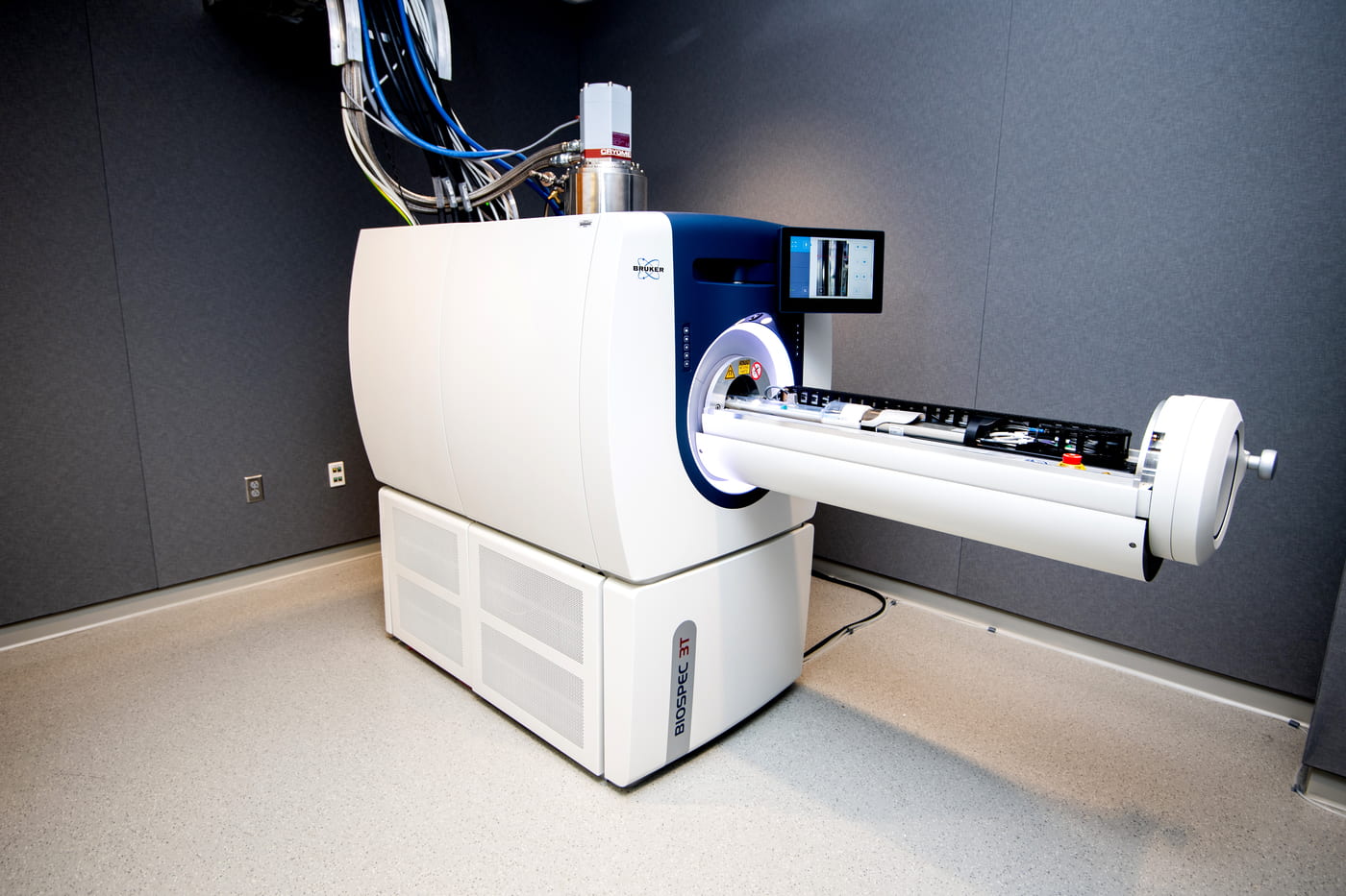
Bruker BioSpec 3T MRI
NU user: $30/hr; Academia: $150/hr; Industry: $200/hr
Automated animal positioning
Multimodal rat/mouse cradle
Two body volume coils for rat/mouse
SAII 1030 monitoring/gating module
Localized MR Spectroscopy
Prerequisite: Bioraft High Magnetic Fields Safety Training
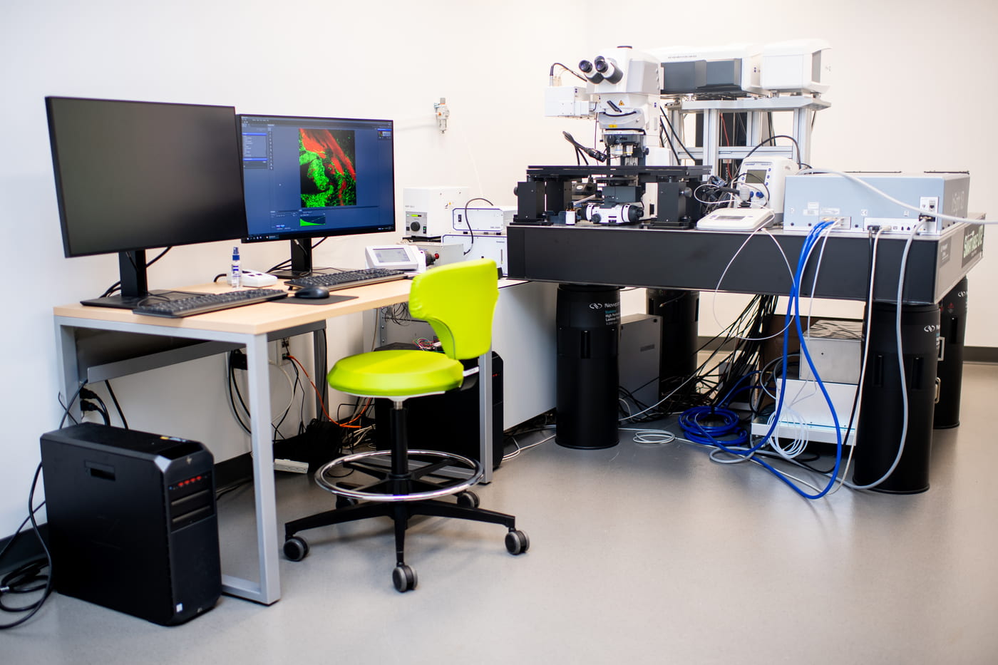
Zeiss LSM 880 NLO (Upright)
NU user: $55/hr; Academia: $75/hr; Industry: $95/hr
Laser line: 405, 458, 488, 514, 561, 594, 640 nm
NIR Laser: tunable line (680-1300 nm); fixed line -1045 nm
Objective: 10×, 20×, 20×, 40×, 20×-CLARITY
Multi-photon imaging, Spectral Unmixing
Prerequisite: Bioraft Laser Safety Training Part I and II
Our facility
Interdisciplinary Science and Engineering Complex
ISEC 080, 805 Columbus Ave, Boston, MA, 02115